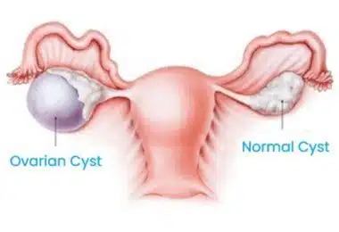
Ovarian Cyst
November 15, 2022
Invasive Diagnostic Methods
November 15, 2022Ovarian Cyst
November 15, 2022Invasive Diagnostic Methods
November 15, 2022
What Is Prenatal Diagnosis (Diagnosis Before Birth)?
Prenatal diagnosis, as a general concept, can be described as the identification of problems of the fetus. The diagnosis of fetal abnormalities or diseases, as a promising field, has become of wide interest, especially due to the latest advances in engineering science and medicine. If the disease is curable in utero, it provides timely assistance and, if treatment requires emergency postpartum intervention, this gives a chance to prepare all the necessary conditions in advance. Finally, in situations of incurable diseases, such a diagnosis allows the discussion of possible termination of pregnancy.
How Is Prenatal Diagnosis Performed?
Today, there are many ways to perform prenatal diagnostics. Some of them are performed by methods that do not involve any risk or discomfort to the mother (non-invasive prenatal diagnosis), while others can pose a danger to the mother and fetus (invasive prenatal diagnosis). This article will briefly describe the non-invasive methods that we use to diagnose the fetus in the womb.
Ultrasound examination during pregnancy in prenatal diagnostics (diagnostics in the womb) is performed using an ultrasound device that, by sending sound waves to the patient’s body through its sensors, studies the characteristics of the returning sound. The sound waves that are sent and returned make the tissues distinguishable from each other and thus make it possible to evaluate the organs. Ultrasound during pregnancy is the most frequently used non-invasive diagnostic method. Due to ultrasound examination during pregnancy, you can detect various problems at different stages of pregnancy. For example, with an ultrasound performed at the beginning of pregnancy, we can find out whether the pregnancy occurs inside or outside the uterus (ectopic pregnancy), accurately determine the number of fetuses (single or multiple pregnancy), the presence of heart contractions, whether the gestational age matches with the actual age of the fetus (dating), as well as the presence of gynecological problems (fibroids, cysts, etc.). In addition, in the first trimester of pregnancy, serious structural abnormalities of the fetus can be detected (intracranial, central nervous system abnormalities, large heart and chest abnormalities, anterior abdominal wall defects, kidney, and urinary tract abnormalities, etc.). In later stages of pregnancy, ultrasound can detect most of the structural problems of the baby. An ultrasound examination during pregnancy can also determine whether the development of the fetus in the womb is consistent with its peers (fetal growth restriction or fetal size), and thus makes it possible to plan the pregnancy accordingly. With the help of ultrasound, it is also possible to understand whether there are such placental pathologies as presentation and invasion anomalies (placental ingrowth: placenta accreta, increta, and percreta). A lack of amniotic fluid (oligohydramnios) or an excess of it in the fetus are also conditions that can be discovered by ultrasound. Ultrasound can also predict whether a pregnancy is dangerous in terms of the occurrence of preterm birth by measuring the length of the cervix. This allows taking appropriate measures to prevent preterm birth. Ultrasonography, as described above, is the most commonly used and very useful diagnostic tool for examining the fetus in the womb. Some images of ultrasonography during pregnancy are shown in Figure 1.
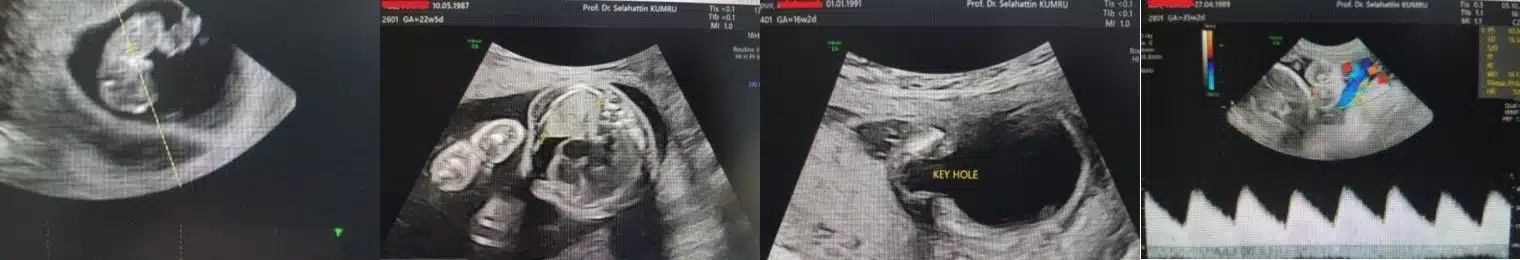
Image-1: Ultrasound during pregnancy (images of some fetal abnormalities that can be recognized with a detailed ultrasound examination). In order: the heartbeat observed in the fetal screening and Color Doppler in the early weeks of pregnancy; the fetus with the accumulation of fluid in the chest cavity diagnosed in the second trimester of pregnancy; the posterior urethral valve diagnosed in the second trimester of pregnancy; the assessment of blood flow in the umbilical cord vein (umbilical artery) of the fetus using Color Doppler in the third trimester of pregnancy.
Color Doppler Ultrasound in Prenatal Diagnosis (the Diagnosis Before Birth)
Color Doppler ultrasound is among the most commonly used invasive methods in pregnancy management. Technically, Color Doppler ultrasound, as well as ultrasound, works by emitting sound waves. It performs the calculation by measuring the speed of moving structures. Thus, you can get an idea of both the properties of the flowing liquid (the density of its contents) and the resistance of the vessels through which it passes.
What Fetus Abnormalities Can Be Diagnosed By Using Color Doppler Ultrasound?
With the help of Color Doppler ultrasound during pregnancy, we can get information about the blood flow in the umbilical cord of the fetus. If there is a lack of essential nutrients supply through the blood flow via the umbilical cord (as in placental insufficiency), the fetus changes the blood flow in its own body (redistribution) to direct incoming oxygen and nutrients to vital organs. This can be detected by Color Doppler screening. When fetal development is delayed due to placental insufficiency, there is an increase in vascular resistance to the blood flow of the umbilical cord. Apart from that, the decrease in the resistance of the blood vessels that feed the brain happens. It prevents brain underdevelopment. This can also be detected by Color Doppler ultrasound, and it helps to find out the cause of the baby’s delays in development. Also, with the help of Color Doppler ultrasound during pregnancy, it is possible to diagnose the presence of anemia in the fetus with great accuracy non-invasively (without any risk involved). In the case of anemia, the flow rate increases because the density of blood circulating in the vessels decreases. By measuring the blood flow rate in the vessels that feed the brain using Color Doppler ultrasound and comparing it with the average blood flow rate of healthy babies, we can understand whether the fetus suffers from anemia or not. In cases of Rh incompatibility or parvovirus infection that cause fetal anemia, doctors have a chance to make a diagnosis even before the baby’s condition worsens, which makes it possible to conduct timely intrauterine intervention (blood transfusion in the womb), which increases the chances of fetal life.